How do you assess for hypertrophic obstructive cardiomyopathy?
Diagnosis – HOCM The diagnosis is made with echocardiography, which will directly visualize the hypertrophied interventricular septum. The ECG in a patient with HOCM will show left ventricular hypertrophy.
Is hypertrophic cardiomyopathy diagnosed?
How Is Hypertrophic Cardiomyopathy Diagnosed? HCM is diagnosed based on medical history (your symptoms and family history), a physical exam, and echocardiogram results. Additional tests may include blood tests, electrocardiogram, chest X-ray, exercise stress test, cardiac catheterization, CT scan, and MRI.
How does a physical exam diagnose hypertrophic cardiomyopathy?
On physical examination, the presence of a harsh crescendo–decrescendo systolic murmur at the lower left sternal border, a mid–late systolic apical murmur or holosystolic apical murmur, and/or paradoxically split S2 should alert clinicians to the possibility of HCM.
How cardiomyopathy is diagnosed?
Diagnosing cardiomyopathy The diagnosis of cardiomyopathy is often clear from an individual’s descriptions of his or her symptoms, the results of a physical examination, and the results of a chest x-ray, echocardiogram, and electrocardiogram. Occasionally, a test called an endomyocardial biopsy is necessary.
What is the difference between hypertrophic cardiomyopathy and hypertrophic obstructive cardiomyopathy?
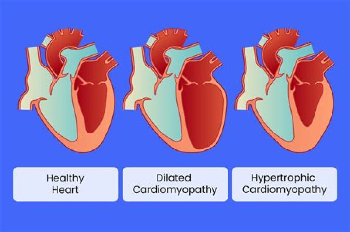
As a result, the thicker wall may block blood flow out of the heart. This is called obstructive hypertrophic cardiomyopathy. If there’s no significant blocking of blood flow, the condition is called nonobstructive hypertrophic cardiomyopathy. However, the heart’s main pumping chamber (left ventricle) may become stiff.
Does ECG show hypertrophic cardiomyopathy?
Test based on electrocardiograms (ECG) that record the heart electrical activity can help in early detection of patients with hypertrophic cardiomyopathy (HCM) where the heart muscle is partially thickened and blood flow is (potentially fatally) obstructed.
Does hypertrophic cardiomyopathy show up on ECG?
The classic ECG finding in hypertrophic obstructive cardiomyopathy is large dagger-like “septal Q waves” in the lateral — and sometimes inferior — leads due to the abnormally hypertrophied interventricular septum. Criteria for left ventricular hypertrophy is usually present.
What causes hypertrophic obstructive cardiomyopathy?
Hypertrophic cardiomyopathy is usually caused by abnormal genes (gene mutations) that cause the heart muscle to grow abnormally thick. In most people with hypertrophic cardiomyopathy, the muscular wall (septum) between the two bottom chambers of the heart (ventricles) becomes thicker than normal.