Why is it important to use neuronavigation in brain stimulation research?
Neuronavigation systems have become extensively used in the operative management of brain tumors and offer a number of advantages to the surgeon. Precise planning of the incision and craniotomy and the identification of small subcortical lesions are some of the principal benefits.
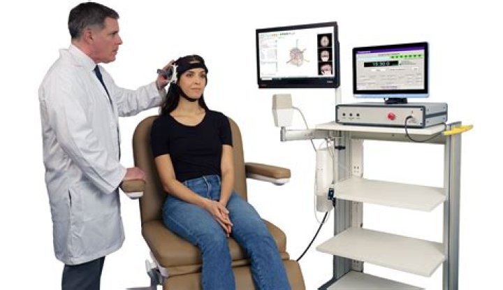
How does neuronavigation work?
Neuronavigation provides a precise surgical guidance by referencing this coordinate system of the brain with a parallel coordinate system of the three-dimensional image data of the patient that is displayed on the console of the computer-workstation so that the medical images become point-to-point maps of the …
What is aneurysm obliteration?
Efficacy (long-term success or effectiveness of the treatment) is measured by evidence of aneurysm obliteration (failure to be demonstrated by conventional or noninvasive angiography), without evidence of recanalization (any blood flow into the aneurysm) or recurrence (reappearance).
Can a 2 mm brain aneurysm rupture?
However, many experienced neurosurgeons and endovascular therapists report that most ruptured aneurysms encountered in practice are small. As seen in our study, aneurysms smaller than 2 mm can also result in an SAH and constituted 7% of ruptured aneurysms in our short experience.
Can brain aneurysm go away?
Aneurysms develop over a lifetime,” he says. “Another is that an aneurysm can disappear or heal itself. This is very rare and only happens in aneurysms that are considered benign because the flow of blood is so slow it eventually forms a clot and seals off the bulge.”
What is the purpose of tractography?
Magnetic resonance diffusion tractography is a method for identifying white matter pathways in the living human brain. These pathways form the substrate for information transfer between remote brain regions and are therefore central to our understanding of function in both the normal and diseased brain.
How does diffusion tractography work?
Diffusion tractography uses non-invasive brain imaging data to trace fibre bundles in the human brain in vivo. This raises immediate possibilities for clinical application but responsible use of this approach requires careful consideration of the scope and limitations of the technique.
How long do you stay in the hospital after a craniotomy?
During the procedure. A craniotomy generally requires a hospital stay of 3 to 7 days. You may also go to a rehabilitation unit for several days after your hospital stay. Procedures may vary depending on your condition and your doctor’s practices.
Can a spinal aneurysm be detected on an MRI?
Unlike brain aneurysms, spinal aneurysms tend to be very small and are almost never recognized on an MRI. A spinal angiogram is usually necessary for diagnosis. It is also a fact that many spinal angiograms are unfortunately inferior in quality.
What is an anterior spinal arterial aneurysm?
Spinal Arterial Aneurysm. The vast majority are fusiform, likely dissecting, aneurysms of the proximal intradural portion o the radiculomedullary or radiculopial artery. The former is an artery which supplies the anterior spinal arterial system, the latter supplies posterior spinal system.
What is the prognosis of spinal aneurysm ischemia?
When aneurysms involve dominant radiculomedullary artery, cord ischemia may result from aneurysm thrombosis or distal embolus. Prognosis in such cases is much worse, as dictated by anatomy. Occlusion of, or embolism into the posterior spinal arteries is usually very well tolerated.
What is the clinical presentation of a ruptured spinal cord aneurysm?
Clinical presentation, when ruptured, is perfectly explained by anatomy. Rupture presents with sudden onset of back pain. Location of initial pain usually corresponds to location of aneurysm, however it tends to quickly spread, as blood products do. If enough bleeding takes place to cause cord compression, the appropriate symptoms follow.